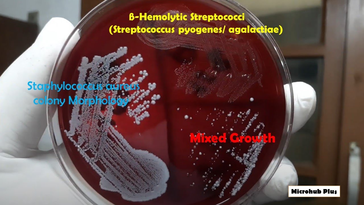
Understanding Blood Agar Plate Hemolysis: A Quick Guide What Is Hemolysis on Blood Agar Plates? Explained Decoding Hemolysis Patterns on Blood Agar Plates Blood Agar Plate Hemolysis: Key Insights & Uses How to Identify Hemolysis on Blood Agar Plates

Blood agar plates are essential tools in microbiology, used to identify and differentiate bacteria based on their ability to lyse red blood cells (RBCs). This process, known as hemolysis, creates distinct patterns that help classify bacterial species. Understanding hemolysis on blood agar plates is crucial for clinical diagnostics, research, and educational purposes. In this guide, we’ll explore what hemolysis is, how to decode its patterns, and its practical applications.
What Is Hemolysis on Blood Agar Plates? Explained

Hemolysis refers to the breakdown of red blood cells, resulting in the release of hemoglobin. On blood agar plates, bacteria produce enzymes or toxins that cause hemolysis, creating visible zones around colonies. This phenomenon is categorized into three types: alpha-hemolysis, beta-hemolysis, and gamma-hemolysis. Each type provides valuable insights into the bacterial species being cultured, aiding in identification and diagnosis.
Decoding Hemolysis Patterns on Blood Agar Plates

Alpha-Hemolysis
Alpha-hemolysis appears as a greenish discoloration around the bacterial colony. This occurs due to the partial lysis of RBCs and the oxidation of hemoglobin to methemoglobin. Bacteria like Streptococcus pneumoniae typically exhibit this pattern.
Beta-Hemolysis
Beta-hemolysis is characterized by a clear zone around the colony, indicating complete RBC lysis. This is caused by hemolysins produced by bacteria such as Streptococcus pyogenes. Beta-hemolysis is often associated with pathogenic strains.
Gamma-Hemolysis
Gamma-hemolysis, or non-hemolysis, shows no visible change in the blood agar. The RBCs remain intact, as seen with bacteria like Staphylococcus epidermidis. This pattern suggests the absence of hemolytic activity.
| Hemolysis Type | Appearance | Example Bacteria |
|---|---|---|
| Alpha | Greenish discoloration | *Streptococcus pneumoniae* |
| Beta | Clear zone | *Streptococcus pyogenes* |
| Gamma | No change | *Staphylococcus epidermidis* |

Blood Agar Plate Hemolysis: Key Insights & Uses

Hemolysis patterns are vital for bacterial identification and diagnosing infections. For instance, beta-hemolytic Streptococcus species are often linked to severe infections like strep throat. Additionally, hemolysis tests help differentiate between closely related bacterial species, enhancing accuracy in clinical microbiology.
How to Identify Hemolysis on Blood Agar Plates

Identifying hemolysis involves careful observation of colony morphology and the surrounding agar. Here’s a quick checklist:
- Inspect for color changes (greenish for alpha, clear for beta).
- Measure the zone size around the colony.
- Compare with known hemolysis patterns for accurate identification.
💡 Note: Always use fresh blood agar plates for accurate results, as older plates may yield misleading patterns.
In summary, understanding hemolysis on blood agar plates is a fundamental skill in microbiology. By recognizing alpha, beta, and gamma patterns, you can identify bacterial species efficiently, aiding in clinical diagnostics and research. Whether you’re a student, researcher, or healthcare professional, mastering this technique is invaluable for your work.
What causes beta-hemolysis on blood agar plates?
+Beta-hemolysis is caused by bacterial hemolysins that completely lyse red blood cells, creating a clear zone around the colony.
Can gamma-hemolysis indicate a non-pathogenic bacterium?
+Yes, gamma-hemolysis (no hemolysis) is often associated with non-pathogenic bacteria, though exceptions exist.
Why is alpha-hemolysis greenish in color?
+The greenish color results from the oxidation of hemoglobin to methemoglobin due to partial RBC lysis.
Related: Bacterial Identification Techniques,Clinical Microbiology Essentials,Laboratory Culture Methods


